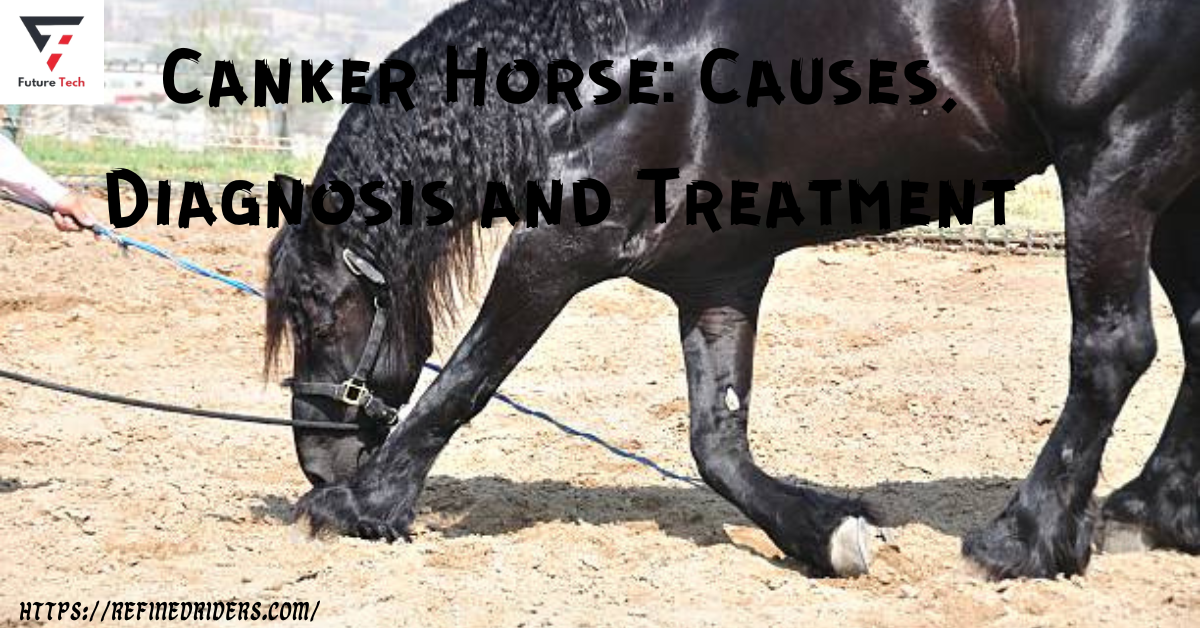
Equine canker is an infectious condition that causes the tissue-producing horns to become chronically larger. Usually starting in the frog, it may spread and infiltrate the surrounding sole, bars, and hoof wall. It can also stay focal. A canker may affect one foot or more than one foot. While it may impact any breed or gender, draft breeds are particularly susceptible to it.
Two imported Warmblood horses that one author (SEO) saw recently had severe canker. There has been a longstanding belief that individuals develop itchiness in moist, humid environments or indolent temperatures. There has been a longstanding belief that individuals develop itchiness in moist, humid environments or indolent temperatures..
However, the exact cause of the disease is still unknown.
Nevertheless, canker is more prevalent in well-groomed horses and animals with regular foot care. As most patients came to his hospital between July and December, one author (JBM) noted that canker was a seasonal occurrence in Florida. Debridements and topical medicine administration, such as antibiotics, astringents, antiseptics, and caustic powders, have been the primary therapies reported in the literature. There is currently no consistently effective therapy for this condition, and the prognosis has always been uncertain…
The Cause of Canker
Bacteria that produce an enlargement of the horn or abnormal keratin production are the causes of horse canker. When the frog’s oxygen supply runs out as a result of this overgrowth, bumps might emerge. There is no evidence to support one notion that canker arose in unsanitary settings. Inadequate hoof-trimming programs are the leading cause of canker development. Horses with cycles longer than eight weeks, as opposed to 4-6 weeks, often exhibit bumps.
An overgrowth of hoof walls results from extending the trimming cycle, which leaves a lot of room for poop, dung, and dirt to settle in.
Clinical Signs
Canker, in its early stages, may be confused for thrush due to its prevalence in amphibians.
I.e., Canker may enter the frog’s horn at any point on its body. Whereas thrush can only enter via the lateral and medial sulci or the animal’s base in the event of a fissure. In contrast to thrush, which causes tissue loss, canker causes tissue to increase. In the first phases, the frog may have a localized region of granulation tissue that is prone to bleeding upon abrading. A light brown or gray tissue will surround this focal spot upon closer examination.
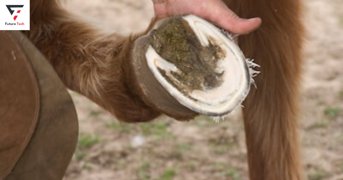
The absence of treatment will result in damage to the frog, the bars, the sole, the palmar/plantar aspect of the foot, and the stratum medium of the hoof wall. The infection causes dyskeratosis, or aberrant keratin formation, which manifests as filamentous horn fronds.2. Many tiny, finger-like papillae of soft, off-white substance that resemble cauliflower are the hallmark of canker.
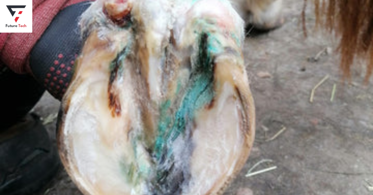
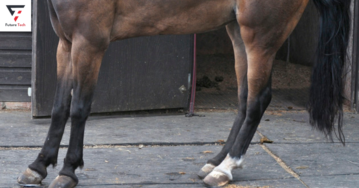
A caseous, white discharge that resembles cottage cheese is the disorder’s defining feature that usually comes with an awful smell, but not always. The horny frog tends to hide the majority of the illness, undermining the frog constantly. When irritated, the damaged tissue may flow readily and may hurt tremendously. Depending on the depth and scope of the infection. There will be varying degrees of lamenessAlthough uncommon, lameness in horses is effectively treatable and diagnosable.
When lameness is present, it usually means that the condition is more severe than just surface excitement and that a proactive approach to treatment is necessary.
Diagnosis
A definitive diagnosis of canker, based on the appearance of the affected horny tissue and an unpleasant stench, may need a biopsy. A biopsy is particularly helpful when the lesions are recurring, do not have a recognizable look, or occur in odd places on the foot. Before performing a biopsy on the superficial necrotic tissue obtained from the periphery of the lesion. It is imperative to exercise caution.
It is essential to biopsy both normal and abnormal tissue.3. An effective biopsy punch is 6 mm. The histological examination of the lesion confirms that it is persistent, hypertrophic, moist frog pododermatitis. Proliferative papillary hyperplasia of the epidermis, dyskeratosis, keratolysis, and ballooning degeneration of the outer layers of the epidermis are its defining characteristics. The stratum germinativum layer of the frog’s epidermis has a diverse variety of bacterial species. 2 Cultures, in general, are unsatisfactory because they usually yield a variety of environmental organisms, including Fusobacterium necrophorum and Bacteroides sp. 3, 4,
Lameness in horses is uncommon, but it is treatable with the appropriate approach.
Treatment
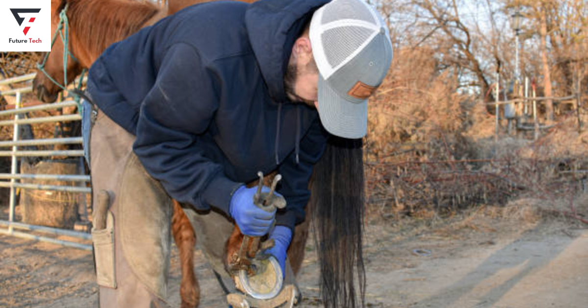
Canker usually has a cautious prognosis. However, these authors have had success lately using the following strategy discontinuing the application of a topical medication on a daily basis until the condition resolves. It is imperative to thoroughly and meticulously debride the afflicted area. General anesthesia or localized anesthetic may be administered to the horse. While it is vertical in order to facilitate the debridement of the injured tissue.By eliminating any exfoliating sole or toe. The hoe’s foot is properly trimmed.
It is essential to use a tourniquet. A cohesive bandage wrapped tightly or with an Esmarch bandage provides firm pressure over the vascular bundles across the abaxial surface of the sesamoids. There are two methods for carrying out debridement. While the horse is under general anesthesia, one author (JBM) employs electric cautery.
In contrast, the other author (SEO) utilizes a sharp hoof knife, a number 12 scalpel blade, and cryotherapy while the animal is upright. Permanent removal of all aberrant tissue until the corium returns to normal.
Separation
There will be an apparent separation between the unusual and normal tissue. It is crucial to avoid removing too much corium as this might impede the cornification process after surgery and lower the caliber and depth of the newly formed sole. To ensure all aberrant tissue is gone, it might be helpful to cut away 1-2 cm of normal tissue from the edges of the incision.
This method of handling the grip swiftly slices through the sole and frog, leaving just a dry eschar. It is possible to lessen the base of the mass by twisting the cautery tip if needed. An analogous approach involves the utilization of a pointed hoof knife for debridement.The afflicted region is cryotherapy-cooled after debridement.
Coolant Spray
While another feasible approach is to use a coolant spray appropriate for electrical circuits to freeze the debrided region, liquid nitrogen has long been the preferred option for this use. The foot’s debrided area will feel pliable and soft. After allowing the affected area to thaw, repeat the freezing procedure until the tissue hardens (often called a hard freeze). Clingy To pack the defect, take four × four sponges and dunk them in a 10% benzoyl peroxide in acetone solution. After that, dust the sponges with a fine powder you made by pulverizing metronidazole pills using a pestle and mortar.
Made of a putty-elastomer composition, the shoe insert ensures that the medication reaches the deepest area of the lesion and reduces the quantity of granulation tissue in significant flaws.The imprinting material should stay within the hoof wall’s bearing surface since doing so will put too much strain on the horse and cause pain. It is then necessary to apply a dry bandage on the foot.
Dry Environment
Maintaining the animal in a dry environment is essential. You may also wear a shoe with a treatment plate, although this approach often makes it challenging to keep the foot as dry as needed. The writers are in favor of bandaging. Standing the horse and applying local anesthetic with laser photoablation or cryotherapy may help treat minor recurrences. The authors raise doubts about the efficacy of systemic antibiotics such as oxytetracycline and chloramphenicol, citing situations where local therapy was sufficient to resolve.4.
SUMMARY
Canker is an infectious disease that affects one or more feet and causes horses’ horns to become more significant over time. It is more common among well-groomed horses and animals that take good care of their feet. The culprit is aberrant keratin synthesis or bacteria that cause the horn to expand, which reduces the frog’s oxygen supply. The frog, bars, sole, palmar/plantar aspect of the foot, and the stratum medium of the hoof wall are all susceptible to canker injury. Lameness, filamentous horn fronds, and dyskeratosis are possible symptoms of the illness.
Horses often develop canker, but it is treatable with a method that requires stopping regular topical therapy. Using either a localized or general anesthetic, debridement of the afflicted region is essential. A hoof knife or tourniquet works well for trimming and debriding the horse’s foot. Following debridement, the damaged area is cryotherapy-cooled and gets daily cleanings and treatments. The authors contend systemic antibiotics are ineffective and stress the need for owner commitment.